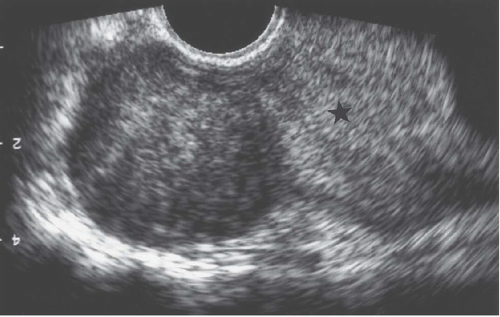
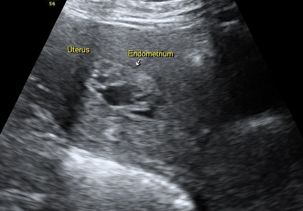
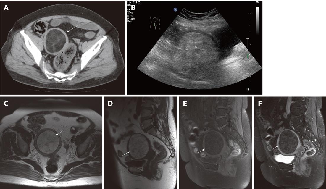
Development, a heterogeneous mass in biology is a tumor with both normal cells and neoplastic cells, which are cells of abnormal growth tissue Heterogeneous masses are called solid tumors and can be cancerous Dr Barry T Kahn from HealthTap explains that heterogeneous masses can be malignant or benignHysterosalpingography reveals enlargement and a distortion of the endometrial cavity by a mural fibroid FIGURE 192 Exophytic fibroid Transvaginal ultrasound reveals a mass that is less echogenic than the adjacent myometrium (star) FIGURE 193 Mural fibroid The uterus is enlarged by a mass of mixed echogenicityIn the first days of menstruation, the heterogeneous structure is typical for the surface of the uterine layer, and the thickness varies from 5 to 8 mm On the third day of the cycle, the process of endometrial transformation develops and its structure acquires good echogenicity
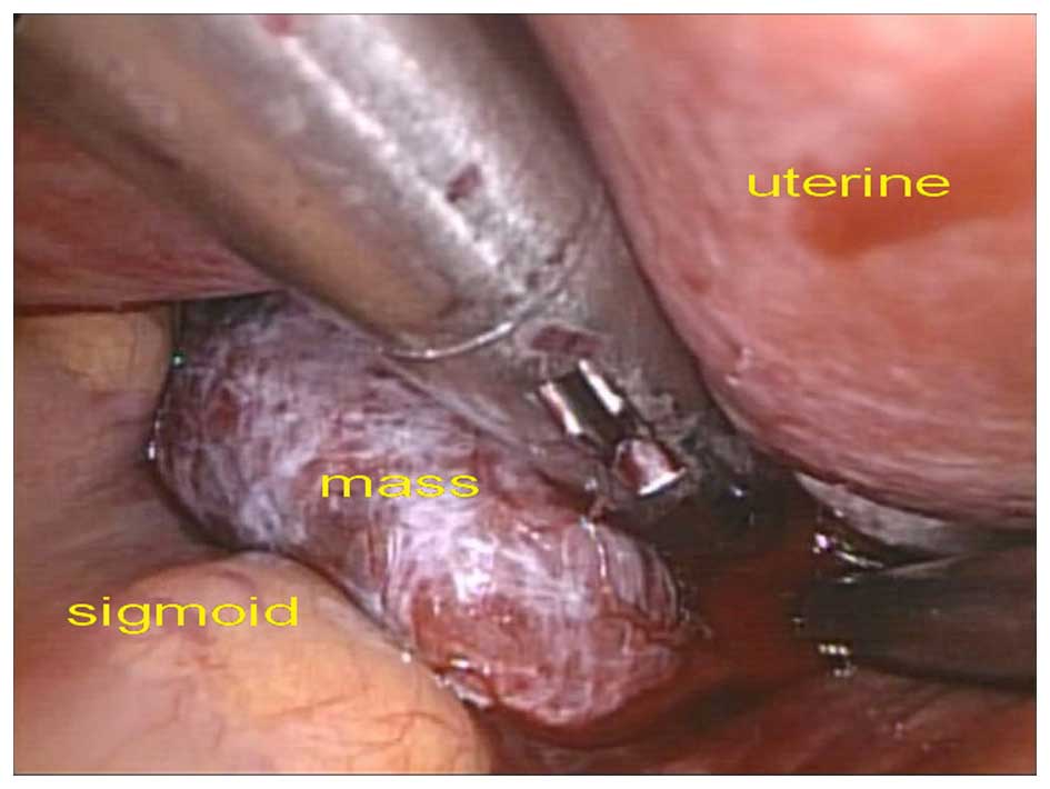
Benign Pelvic Masses Masquerading As Adnexal Cancer During Pregnancy On Ultrasound A Retrospective Study Of 5 Years
Heterogeneous mass in uterus
Heterogeneous mass in uterus-Heterogeneous (and possibly lobulated) cystic mass with internal calcific focus in the right adnexa could be ovarian in etiology;Heterogeneous uterus Heterogenous uterus is a medically sonographic terminology It says how your uterus look under sonographic waves It says how your uterus look under sonographic waves That is not a pathology in itself


Ultrasonography Of Uterine Leiomyomas Sciencedirect
It may mean that a fibroid or cyst was found during the ultrasound Nearly all of these are small and cause no symptoms or problems, but you need to discuss this finding with your doctor What is a heterogeneous uterus?A heterogeneous mass posterior to the uterus generally indicates a fibroid tumor A fibroid is a benign tumor that is non cancerous(b) Sagittal contrastenhanced T1weighted MR image obtained in a 45yearold woman after endometrial polypectomy shows differential enhancement of the cervix and uterus, with a clear demarcation (arrows) between the patchy heterogeneous myometrial enhancement in the cervix and the more homogeneous enhancement in the uterus The vaginal canal is easily
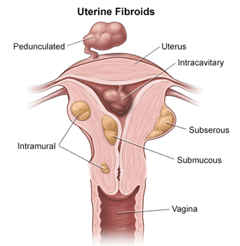
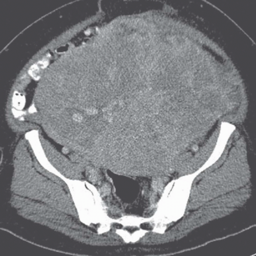
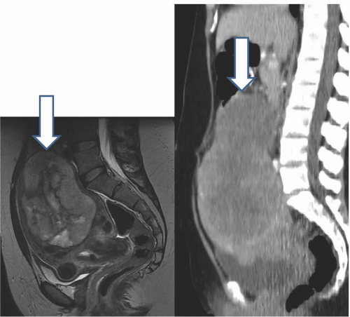
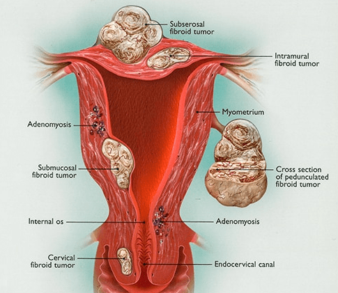
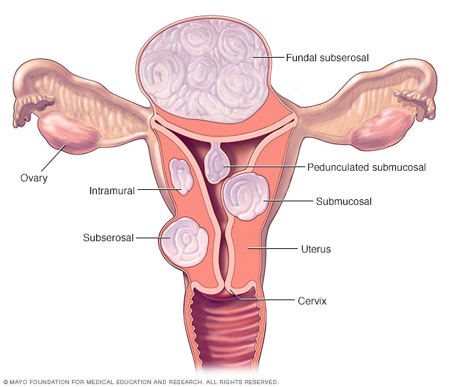
Fibroids usually develop from cells in the wall of the uterus but can also grow beneath the uterine lining When these fibroids expand, they can stretch the endometrium, causing heavy menstrual bleeding and severe pain as the uterus tries to expel the mass Even small fibroids in this location may cause these symptomsThe organ of origin of the mass could not be identified on ultrasonography Contrast enhanced computerised tomography revealed a large heterogeneous, predominantly hypodense mass lesion in the abdomen and pelvis, measuring 169 cm ×Molar pregnancy thickened with multiple small cystic spaces;
Heterogeneous means different textures like blood can look like that mass is another word for spot, nodule, density, and so on Unless you have other issues that are a cause for concern family history ovarian cancer personal history of cancer severe unremitting pain, fever, nausea, vomiting, change in bowelsNonpregnancy related endometrial carcinoma variable appearanceUltrasound Evaluation of Myometrium The myometrium is the muscular tissue of the uterus and the cervix, which encloses the uterine cavity and its lining, the endometrium The myometrium is generally isoechoic (similar to the liver) and homogeneous The myometrial echogenicity, thickness, contour and presence of any mass or cysts are noted
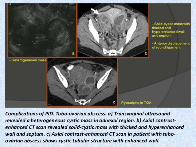


Presentation1 Pptx Radiological Imaging Of Uterine Lesions


Ultrasound Scans Revealed A Large Heterogeneous Mass Occupying The Download Scientific Diagram
Standardized terms to be used when describing ultrasound images of the endometrium and uterine cavity have been suggested by the IETA (International Endometrial Tumor Analysis) group 3, but there remains no standardized terminology for describing ultrasound images of normal or pathological myometrium, or uterine masses 4Most women with uterine fibroids have an enlarged uterus In fact, doctors describe the size of fibroids and their effect on a woman's uterus as they would a pregnancy, such as a 14weeksized fibroid uterus It's not uncommon for a fibroidaffected uterus to grow to the size of a four to fivemonth pregnancy (iii)Large pelvic masses in women may originate from the uterus, cervix, ovaries, fallopian tubes, peritoneum, or retroperitoneum Magnetic resonance (MR) imaging is commonly used in the workup of such lesions Often the diagnosis can be suggested on the basis of tumor location and anatomic landmarks


Uterine Endometrial Cancer Symptoms Staging Treatment Risk Factors


Choriocarcinoma A Rapidly Progressive Unusual Tumor Eurorad
Cancer heterogeneity, long recognized as an important clinical determinant of patient outcomes, was poorly understood at a molecular level Genomic studies have significantly improved our understanding of heterogeneity, and have pointed to ways in which heterogeneity might be understood and defeated for therapeutic effectMy uterus is slightly bulky (95 * 667 * 597 ) cm approx endometrium thickness = mm a well defined heterogenous echotextural intramural >Pelvic ultrasound The findings Uterus measured 16x169x92 cm, Arising from the right uterine body, there is a large mass measuring 72x12x113cm this is somewhat heterogeneous with areas of increas read more Dr Kala Director Women\u0027s Health
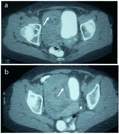


Uterine Lipoleiomyoma A Case Report And Literature Review


Ultrasound Imaging Of Uterine Fibroids Evaluation And Management Chapter 12 Ultrasonography In Gynecology
According to Genes &135 cm and showed heterogeneous irregular enhancing hypodensity within itTeratoma / dermoid cyst is primarily considered Negative for frank invasion of the adjacent bowels, urinary bladder and uterus


The Assessment Diagnosis And Causes Of Endometrial Cancer Empowered Women S Health


When Would You Suspect Fibroid Uterus
The typical finding is a bulky, irregular uterus or a mass in continuity with the uterus Degenerate fibroids may appear complex and contain areas of fluid attenuation Calcification is seen in approximately 4% of fibroids and is typically dense and amorphousUterine mass ICD10CM N858 is grouped within Diagnostic Related Group (s) (MSDRG v380) 742 Uterine and adnexa procedures for nonmalignancy with cc/mcc 743 Uterine and adnexa procedures for nonmalignancy without cc/mcc 760 Menstrual and other female reproductive system disorders with cc/mccBulky uterus with focal lesion welldefined heterogenous mass within measure 34*27cm in maximum dimension with small calcifi foci within,cause colic?


Scielo Brasil Tumors Of The Broad Ligament What And When To Suspect Such Rare Location Tumors Of The Broad Ligament What And When To Suspect Such Rare Location


When Would You Suspect Fibroid Uterus
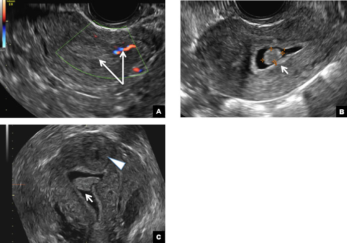
However, if a mass is adjacent to the uterus and is of intermediate or high signal intensity relative to the myometrium on T2weighted images, the differential diagnosis includes both degenerated leiomyoma and extrauterine tumors (benign and malignant) If T1 signal intensity was low or heterogeneous, ADC values obtained at B1000 at 15 THeterogeneous just means that the texture of your uterus is not uniform that can be because of fibroids another common reason is a condition called adenomyosis which isFocal adenomyosis or adenomyoma can be mistaken for a leiomyoma on US 26 Lack of a uterine contour abnormality or mass effect, elliptical shape, increased echogenicity and illdefined margins favor the diagnosis of adenomyoma over leiomyoma 10 Color Doppler US may also aid in distinguishing adenomyoma from leiomyoma on US
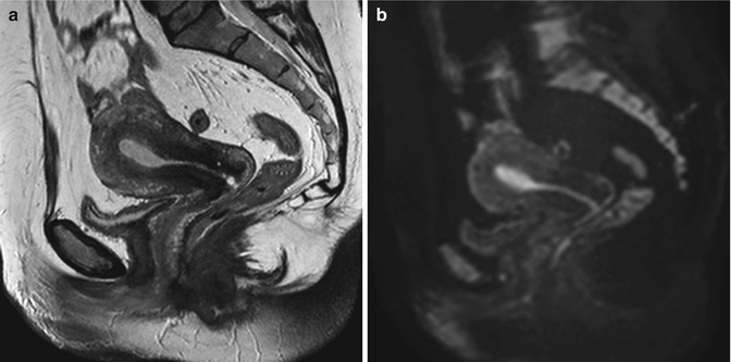


Fibroids Radiology Key


When Would You Suspect Fibroid Uterus
Uterine growths are enlargements, masses, or tumors located in the female womb (uterus) An example of a benign or noncancerous growth is a polyp of the cervix Although uterine fibroids are also benign causes of uterine growths, they can still cause signs and symptoms such as bleeding Dangerous growths of the uterus include cancerous tumorsA Verified Doctor answered A US doctor answered Learn moreUterine arteries, branches of the anterior division of the internal iliac arteries (Fig 1) Yitta et al 13 described a fourth heterogeneous and multifocal enhancement pattern of the entire myometrium (Fig 4 and 10) related to decreased muscle mass and atheromatous changes 2 CT shows a low sensitivity in the assessment of endometrial


Www Bmus Org Static Uploads Resources Uterus Endometrium Common Uncommon Pathologies Bmus York Pdf


Radiology In The Diagnosis Staging And Management Of Gynecologic Malignancies Glowm
Most malignant masses are predominantly hypoechoic, usually with lowlevel echoes or heterogeneity, and well defined as they have pseudocapsules As a malignant mass enlarges, especially if high grade, it becomes more heterogeneous with hypoechoic areas of internal necrosis and increased flow on color and power Doppler imagingThe report says a large, lobulated heterogeneous echo pattern mass is seen in the right side of the uterus, shifting the uterus towards left It measures 131*128*102 cm Bilateral ovaries are not seen distinctly No free fluid seen What is this disease?They are typically large, heterogeneous masses containing areas of hemorrhage They usually have more illdefined, irregular margins, compared to benign uterine leiomyomas Combined grayscale and color Doppler US may help distinguish uterine leiomyosarcoma from leiomyoma Adenomyosis is the ectopic endometrial tissue within the myometrium


Answer To What S Your Diagnosis Previous Page Annals Of Saudi Medicine


Malignant Mixed Mullerian Tumors Of The Uterus Sonographic Spectrum Lee 12 Ultrasound In Obstetrics Amp Gynecology Wiley Online Library
A pelvic mass is an enlargement or swelling in the pelvic region Most pelvic masses are discovered during routine gynecologic or physical examinations Pelvic masses may originate from either the gynecologic organs, such as the cervix, uterus, uterine adnexa, or from other pelvic organs, such as the intestines, bladder, ureters, and renal organsAnswer 'Heterogenous uterus' is a description used to describe the appearance of the uterus after an ultrasound exam is done All that this means is that the ultrasound appearance of the uterus is not totally uniform The two most common causes of heterogenous uterus are uterine fibroids, which are benign muscular growths in the uterine wall, andAdnexal/Pelvic Mass (Ovarian Mass, Ovarian Cyst) 1 Epidemiology, Signs and Symptoms In the United States, the diagnosis of an adnexal or pelvic mass will occur in five to ten percent of women in


Cross Sectional Imaging Of Acute Gynaecologic Disorders Ct And Mri Findings With Differential Diagnosis Part Ii Uterine Emergencies And Pelvic Inflammatory Disease Insights Into Imaging Full Text


Effect Of Magnetic Resonance Imaging Characteristics On Uterine Fibroi Rmi
A, Axial T1weighted (333/11) image shows a mass with hyperintensity in the uterus B, Sagittal T2weighted (1800/80) image shows a hyperintense mass (M) with minimal heterogeneity in the uterus A thin hypointense rim is seen to surround the mass and was confirmed to be the compressed myomatous tissue (pseudocapsule)Findings are usually associated with an enlarged uterus;Cancer in the wall of the uterus (where I assume this is located) is rare, and fibroids are very common On ultrasound, fibroids usually appear heterogeneous (not smooth and plain) and may be complex Perhaps there is something about the ultrasound appearance that makes this mass unusual, but I would guess that it's just another fibroid


When Would You Suspect Fibroid Uterus


Scielo Brasil Tumors Of The Broad Ligament What And When To Suspect Such Rare Location Tumors Of The Broad Ligament What And When To Suspect Such Rare Location
It accounts for 5% of all uterine corpus malignancies and is found in postmenopausal women Microscopically it shows an admixture of carcinomatous and sarcomalike elements, resulting in a characteristic biphasic appearance It appears as a large, soft, polypoid mass involving the endometrium and myometriumA heterogeneous uterus is a term used to describe the appearance of the uterus after an ultrasound is conducted It simply means that the uterus is not totally uniform in appearance during the ultrasound According to MedHelp, there are two common causes of a heterogeneous uterus uterine fibroids and adenomyosisA, Sagittal T2weighted pelvic MR image shows heterogeneous ovoid mass arising from anterior cervix (arrowheads), caudal to uterine corpus This example underscores some challenges in imaging leiomyosarcoma because features overlap with features of leiomyomas (eg this mass is ovoid and well defined, features that are more typical for benign


Benign Pelvic Masses Masquerading As Adnexal Cancer During Pregnancy On Ultrasound A Retrospective Study Of 5 Years


Ultrasonography Of Uterine Leiomyomas Sciencedirect
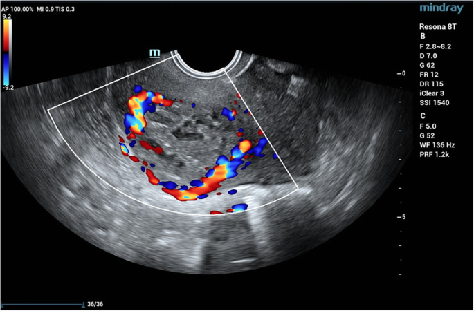
Heterogeneous appearing myometrium Endometrial echo complex is normal dominant follicle upper pole right ovary Normal appearing left ovary The right ovary measure 45 x 24 x 21 cm and the left ovary measures 31 x 17 x 17 You have a small fibroid in your uterus of size 2x1 9x15 It's a small one Don't worryThe ultrasound is from a 70 year old post menopausal female who presents with an enlarged uterus The endometrial stripe is enlarged and is filled with fluid and an enhancing soft tissue mass consistent with an endometrial carcinoma Note blood flow as depicted by Doppler exam (c) characterizing the soft tissue as tumor rather than a clotA) Endometrial polyp INCORRECT Endometrial polyps on ultrasonography appear as focal echogenic (hyperechoic) masses or as nonspecific endometrial thickening 1 Color Doppler often demonstrates a vascular stalk, which is a nonspecific finding that also can be seen in submucosal fibroids and endometrial cancer 2 On sonohysterography (SHG), endometrial polyps typically


Center For Uterine Fibroids


Types Of Fibroids Tumors Houston Fibroids
A hypoechoic mass is an area on an ultrasound that is more solid than usual tissue It can indicate the presence of a tumor, but many times these masses are benign (noncancerous) Because earlyHeterogeneously hypoechoic mass lesion measuring 61x 38 cm seen involving the lower body of uterus and andRecent gestational state (delivery) endometritis;


Effect Of Magnetic Resonance Imaging Characteristics On Uterine Fibroi Rmi


Ultrasonography Of Uterine Leiomyomas Sciencedirect
A heterogeneous mass posterior to the uterus generally indicates a fibroid tumor A fibroid is a benign tumor that is non cancerous What is T2 signal in MRI?Heterogeneous refers to a structure with dissimilar components or elements, appearing irregular or variegated For example, a dermoid cyst has heterogeneous attenuation on CT It is the antonym for homogeneous, meaning a structure with similar components Heterogenous refers to a structure having a foreign originOn the cut surface of the uterus, the lesion was a well circumscribed, intramural, serous fluidfilled cystic mass in the uterine fundus having continuity with the endometrium (Figure 3) On microscopic examination, the cystic masses in the myometrium and the right ovary were seen to be lined by tubaltype epithelium consistent with the


Bulky Uterus It S Symptoms Causes And Treatment Medicover Fertility


When Would You Suspect Fibroid Uterus
Objective The purpose of this presentation is to show the imaging findings of the common and uncommon variants of adenomyosis as seen on sonography and magnetic resonance imaging (MRI) Methods A 3Intrauterine blood clot heterogeneous endometrium with no vascularity;


Differential Diagnosis For Female Pelvic Masses Intechopen


Differential Diagnosis For Female Pelvic Masses Intechopen


Radiological Appearances Of Uterine Fibroids Uterine Fibroids Fibroids Hysterectomy
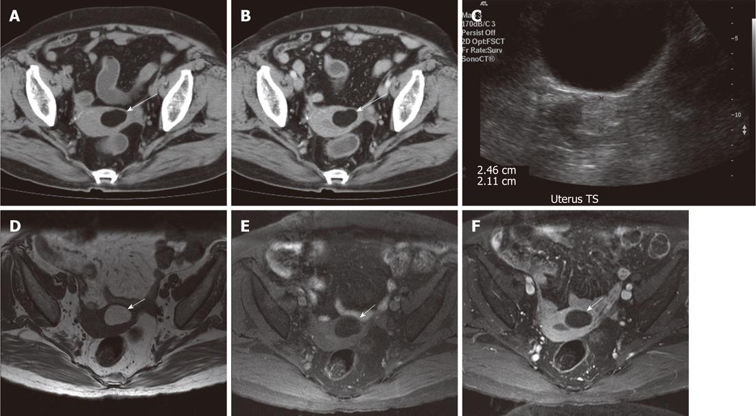


Diagnostic Challenge Of Lipomatous Uterine Tumors In Three Patients


Bulky Uterus It S Symptoms Causes And Treatment Medicover Fertility


Diseases Of The Uterus Radiology Key


Ultrasonography Of Uterine Leiomyomas Sciencedirect
/enlarged-uterus-signs-symptoms-complications-4174349_FINAL_640px-5bc5fb96c9e77c005123488f.png)


Signs And Symptoms Of An Enlarged Uterus


Q Tbn And9gcqcvdfxpgzlf7jt5svsem9fepvvbrprlzbnav4iiia3pcgibjxu Usqp Cau


Ultrasound Image Showing The Bulky Uterus With Mass Lesion Inside The Download Scientific Diagram


42 Year Old Woman With Abnormal Uterine Bleeding Mdedge Obgyn


3


Transvaginal Sagittal Uterine Image Displaying Globular Uterine Enlargement And Heterogeneous Myometrial Texture Uterine Wall Thickening Can Show Anteroposterior Ppt Download


Diseases Of The Uterus Radiology Key
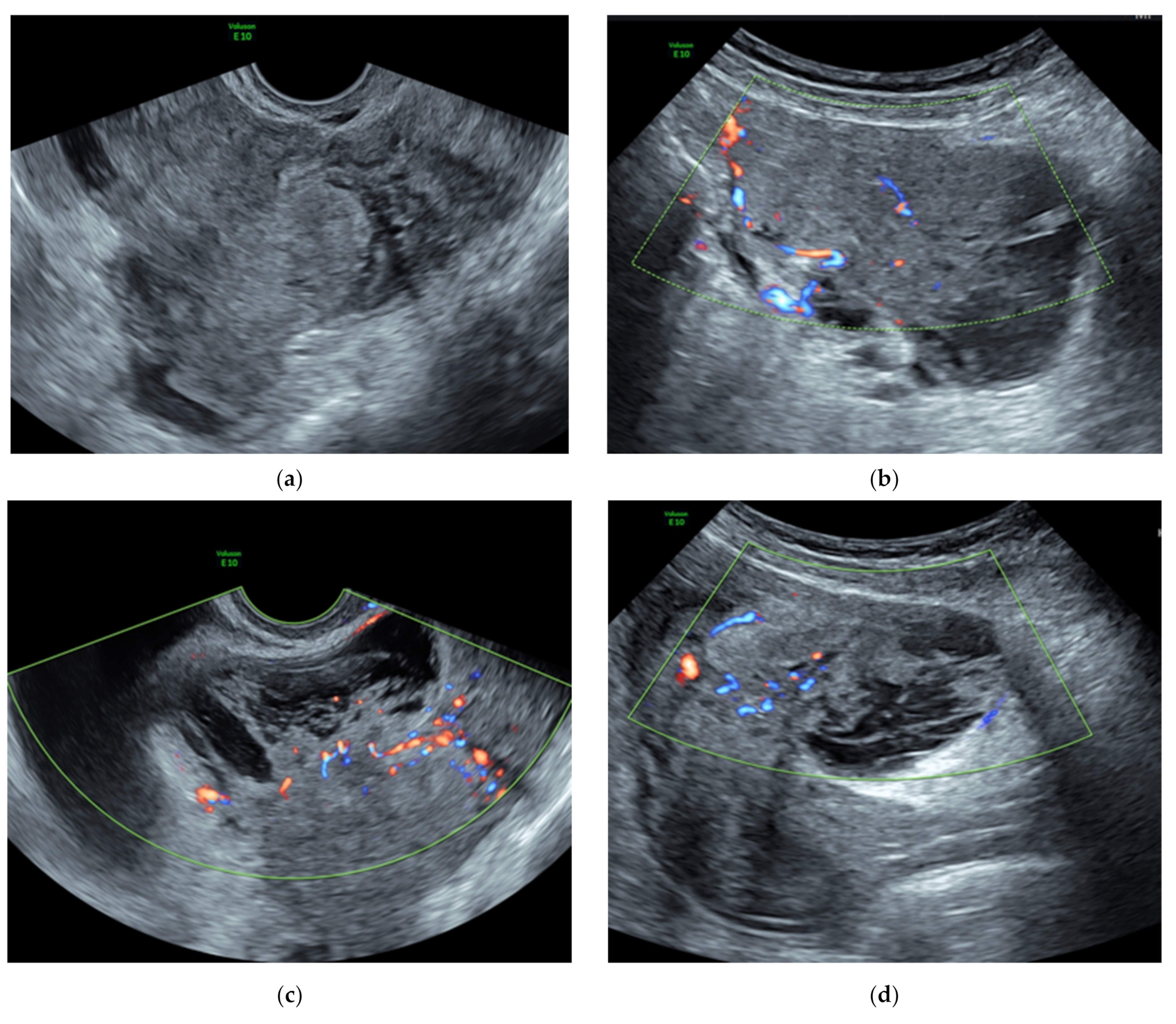


Ijerph Free Full Text The Management Of The Cotyledonoid Leiomyoma Of The Uterus A Narrative Review Of The Literature Html
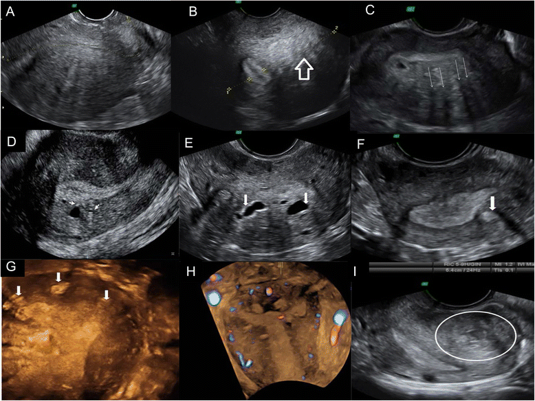


Adenomyosis In Infertile Women Prevalence And The Role Of 3d Ultrasound As A Marker Of Severity Of The Disease Reproductive Biology And Endocrinology Full Text


Computed Tomography Ct Of The Abdomen And Pelvis Demonstrating Solid Download Scientific Diagram


Table 1 From Ultrasonography Evaluation Of Pelvic Masses Semantic Scholar


Radiological Category Genitourinary Principal Modality 1 Ultrasound Principal


Huge Mass With Uterus And Bilateral Ovaries Fig 2 Ultrasound Showing Download Scientific Diagram
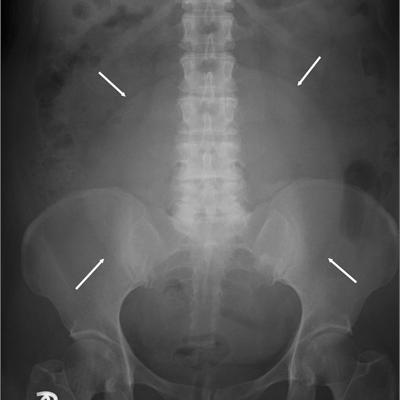


Large Pelvic Mass Mdedge Family Medicine


A Heterogeneous Adnexal Mass Heterogeneous Adnexal Mass Adjacent To Download Scientific Diagram
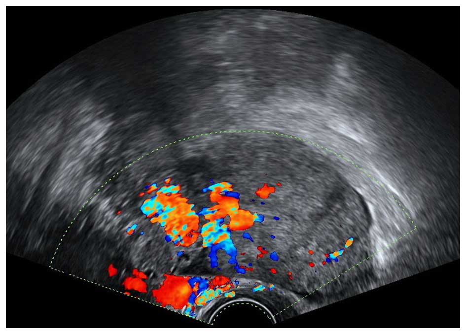


Benign Pelvic Masses Masquerading As Adnexal Cancer During Pregnancy On Ultrasound A Retrospective Study Of 5 Years


Abnormally Thickened Endometrium Differential Radiology Reference Article Radiopaedia Org


Ultrasonography Of Uterine Leiomyomas Sciencedirect


Uterine Mass After Caesarean Section A Report Of Two Cases Bmc Pregnancy And Childbirth Full Text


42 Year Old Woman With Abnormal Uterine Bleeding Mdedge Obgyn


When Would You Suspect Fibroid Uterus


Scielo Brasil Ultrassonografia Nas Massas Anexiais Aspectos De Imagem Ultrassonografia Nas Massas Anexiais Aspectos De Imagem


Diseases Of The Uterus Radiology Key


Terms Definitions And Measurements To Describe Sonographic Features Of Myometrium And Uterine Masses A Consensus Opinion From The Morphological Uterus Sonographic Assessment Musa Group Van Den Bosch 15 Ultrasound
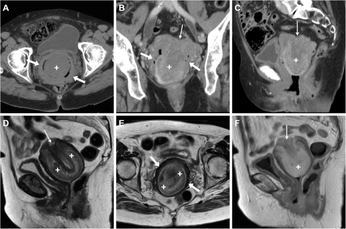


Cross Sectional Imaging Of Acute Gynaecologic Disorders Ct And Mri Findings With Differential Diagnosis Part Ii Uterine Emergencies And Pelvic Inflammatory Disease Insights Into Imaging Full Text
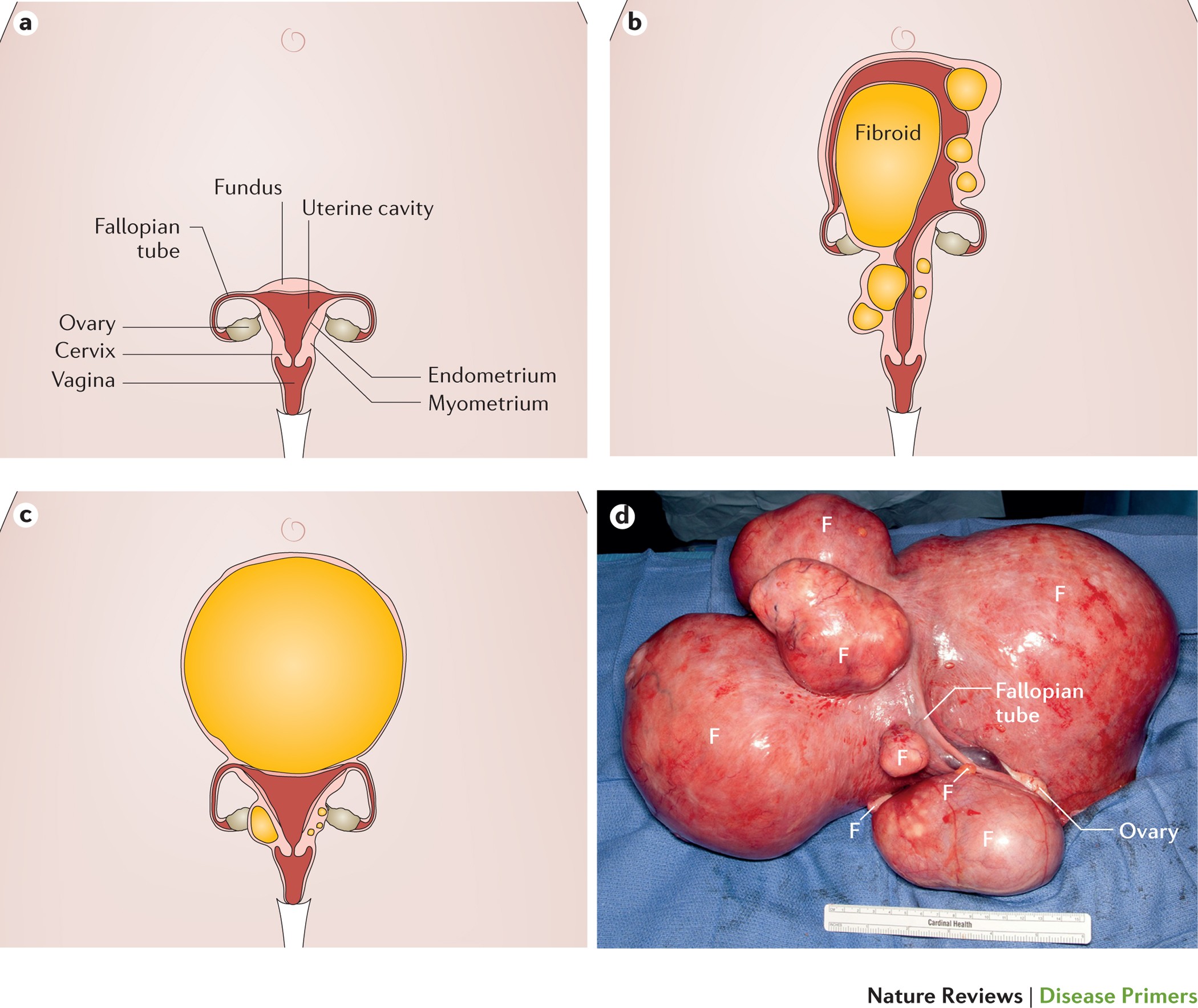


Uterine Fibroids Nature Reviews Disease Primers


Heterogeneous Uterine Mass With Prominent Cystic Lesions Within The Download Scientific Diagram
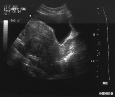


Uterine Leiomyoma Fibroid Imaging Overview Radiography Computed Tomography


Non Puerperal Uterine Inversion
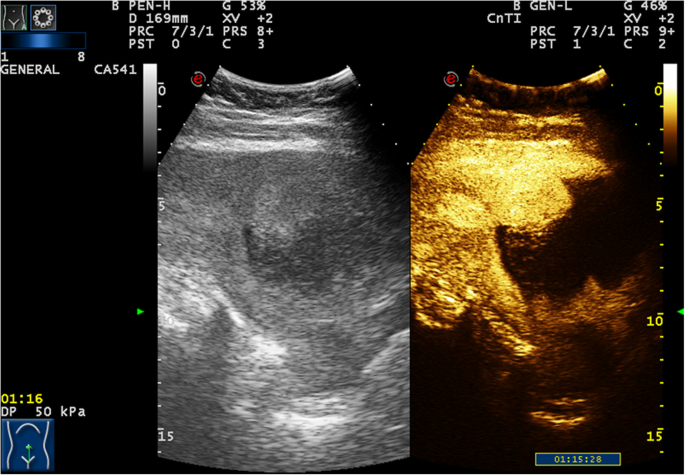


Uterine Mass After Caesarean Section A Report Of Two Cases Bmc Pregnancy And Childbirth Full Text


Q Tbn And9gctchbr5bjpyk1lxrnxpdi4 Tziyt9oyndy4 Clvjjlsi6mr7rmp Usqp Cau


Malignant Mixed Mullerian Tumor Of The Uterus Radiology Case Radiopaedia Org


Ultrasonography Of Uterine Leiomyomas Sciencedirect


Diagnostic Challenge Of Lipomatous Uterine Tumors In Three Patients


Pelvic Pain Overlooked And Underdiagnosed Gynecologic Conditions Radiographics


Ultrasound Evaluation Of Myometrium Obgyn Key


Adenomyosis Common And Uncommon Manifestations On Sonography And Magnetic Resonance Imaging Chopra 06 Journal Of Ultrasound In Medicine Wiley Online Library


Hypoechoic Mass In The Liver Breast Kidney And More


Q Tbn And9gctslmjxpgtqtgbl1uxknivh3sumrgnywxebtsjy6v3t Yjuo6ps Usqp Cau


Ultrasonography Of Uterine Leiomyomas Sciencedirect


Ultrasonography Of Uterine Leiomyomas Sciencedirect


Ct Scan Found Heterogeneous Mass With Solid And Cystic Parts In Uterus Download Scientific Diagram


Symptoms Of Uterine Cancer Vs Fibroids


Ultrasonography Of Uterine Leiomyomas Abstract Europe Pmc


A Pelvic Ultrasound Shows A Well Circumscribed Lobulated Download Scientific Diagram


Common Gynaecological Problems And Their Treatment Dr Sita Sharma Mbbs Md Obstetrics And Gynecology


When Would You Suspect Fibroid Uterus


Ultrasonography Of Uterine Leiomyomas Sciencedirect


Transvaginal Ultrasonography Revealed A Complex Heterogeneous Tumor In Download Scientific Diagram


When Would You Suspect Fibroid Uterus


Ultrasound Scans Revealed A Large Heterogeneous Mass Occupying The Download Scientific Diagram


When Would You Suspect Fibroid Uterus


Uterine Fibroids Symptoms And Causes Mayo Clinic


Intravenous Leiomyomatosis With Right Atrium Extension In Two Patients A Case Report


Ultrasound Characteristics Of Endometrial Cancer As Defined By International Endometrial Tumor Analysis Ieta Consensus Nomenclature Prospective Multicenter Study Epstein 18 Ultrasound In Obstetrics Amp Gynecology Wiley Online Library


The Assessment Diagnosis And Causes Of Endometrial Cancer Empowered Women S Health


Ultrasonography Of Uterine Leiomyomas Sciencedirect
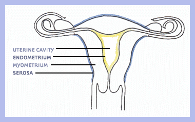


Center For Uterine Fibroids


Ultrasonography Of Uterine Leiomyomas Sciencedirect
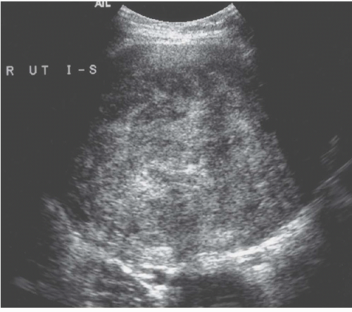


Diseases Of The Uterus Radiology Key
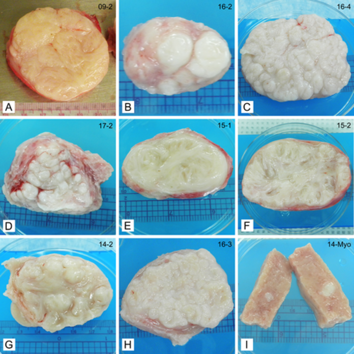


Recognizing Heterogeneity In Fibroids Could Expand Treatment Options Duke Health Referring Physicians


Uterine Corpus Uterus Leiomyoma


Scielo Brasil Ultrassonografia Nas Massas Anexiais Aspectos De Imagem Ultrassonografia Nas Massas Anexiais Aspectos De Imagem
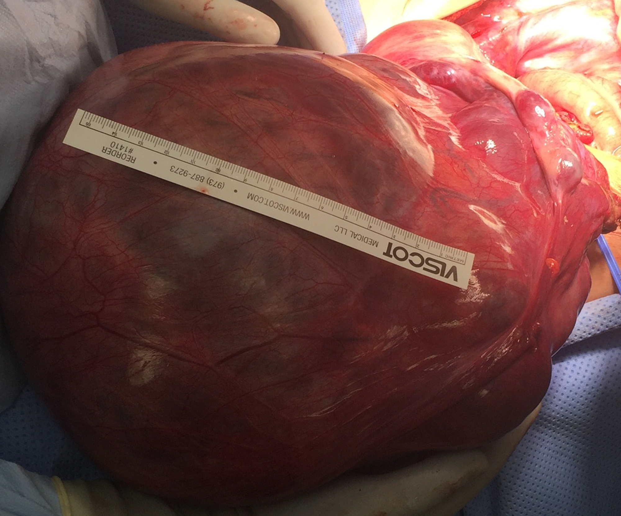


Cureus Myomatous Erythrocytosis Syndrome Case Report And Review Of The Literature


Cureus Polypoid Adenomyoma Of The Uterus


Doppler Ultrasound In Gynaecology Chapter 16 Gynaecological Ultrasound Scanning


When Would You Suspect Fibroid Uterus


Magnetic Resonance Imaging Of Non Puerperal Complete Uterine Inversion Iranian Journal Of Radiology Full Text


J Clin Gynecol Obstet


0 件のコメント:
コメントを投稿